| Can shoulder
dystocia be anticipated accurately?
The answer to this question by the vast majority of experts in obstetrics is “No”. This is confirmed by:
The ACOG Bulletin 40 (2002, reaffirmed 2015) which says “Shoulder dystocia is most often an unpredictable and unpreventable obstetric emergency.
The ACOG publication Neonatal Brachial Plexus Palsy (2014), p. 17: “Risk factors for shoulder dystocia are not reliable predictors for its occurrence.”
Williams Obstetrics (25th Edition, 2014): “Identification of individual instances [of shoulder dystocia] before the fact has proven to be impossible.…. Most cases of shoulder dystocia cannot be accurately predicted or prevented.”
In the past, there have been physicians who have claimed that shoulder dystocia could be predicted. Hassan (1988) stated,
"In the majority of cases shoulder dystocia can be anticipated. Risk factors include maternal obesity, diabetes, preeclampsia, prolonged gestation, and fetal macrosomia. A male infant is at a greater risk for macrosomia and dystocia."
O'Leary, in his 1992 book, Shoulder Dystocia and Birth Injuries, concurred.
However, this has been an overwhelmingly minority opinion. The vast majority of obstetricians, including those who have done the most work on shoulder dystocia and brachial plexus injuries, have concluded that it is impossible with any degree of certainty to predict in which deliveries shoulder dystocia will occur. The key issue involved is "certainty". As will be shown, there are multiple "risk" factors for shoulder dystocia. Mothers and babies having these risk factors are, in an absolute sense, more likely than mothers and babies without these factors to experience shoulder dystocia. But whether the predictive value of such factors as currently measured is high enough to be useful clinically, that is, to justify changes in labor management in hopes of avoiding shoulder dystocia, is what is at issue.
Moreover, as with most statistical questions in medicine, the predictability of shoulder dystocia has to be looked at from two points of view:
Sensitivity: Are the risk factors associated with shoulder dystocia able to accurately identify most babies who will experience a shoulder dystocia at birth?
Positive predictive value: What percentage of mothers and babies having these risk factors will, in fact, experience shoulder dystocia?
In the case of shoulder dystocia, its infrequent rate of occurrence (0.5%-1.5%) and the low positive predictive value risk factors for it have severely impeded the ability of obstetricians to utilize such information to advantageously alter clinical care.
The medical literature confirms this overwhelmingly.
Resnick (1988), discussing the ability of obstetricians to predict when shoulder dystocias will occur, stated that "the diagnosis [of shoulder dystocia] will often be made only after delivery of the fetal head."
Geary (1995) reported that when all antenatal risk factors for shoulder dystocia were taken into account, the positive predictive value was less than 2% for individual factors and less than 3% when multiple factors were combined.
Lewis (1998) noted that only 25% of shoulder dystocia cases had at least one significant risk factor.
Gherman (2002), among current leaders in the study of shoulder dystocia, has said the following:
“Most of these preconception and prenatal risk factors have extremely poor positive predictive values and therefore do not allow the obstetrician to accurately and reliably predict the occurrence of shoulder dystocia.”
The obstetrical literature contains many other articles which share this point of view.
The general consensus in obstetrics is that both the sensitivity and positive predictive value for predicting shoulder dystocia is far too low to justify obstetrical interventions in hopes of avoiding it.
However, the above dictum has been challenged, particularly for shoulder dystocia with brachial plexus injury. There has been work by Emily Hamilton et al in Montreal using statistical methods to estimate the risk of shoulder dystocia with brachial plexus injury. The assessment of risk is based on the size of both the baby and the mother. This work indicates that it is possible to identify a small subgroup with very elevated risk of shoulder dystocia with brachial plexus injury, where the tradeoff between potential prevention and unnecessary intervention matches or exceeds the results using the ACOG intervention criteria.
The original algorithm evaluated the following factors: previous vagina birth, mother’s height and weight, gestational age, and estimated fetal weight. In a 2006 paper, Dyachenko and Hamilton showed that their algorithm was able to detect 50.7% of the cases of shoulder dystocia with some brachial plexus injury along with a false positive rate of only 2.7%. In a second study published in 2012 (Daly, 2012), the clinicians employed a similar algorithm prospectively, in just under 9000 deliveries from two New Jersey hospitals. Use of the algorithm resulted in a lowering of the rate of shoulder dystocia by 56.8% while not at all increasing the rate of primary cesarean sections.
Whether or not the Hamilton algorithm will change the current consensus in obstetrics that shoulder dystocia is unpredictable awaits further verification.
� Categories of risk factors
The risk factors for shoulder dystocia can generally be divided into three categories:
Preconceptual — before pregnancy
Antepartum — during pregnancy
Intrapartum — during labor and delivery
A. Preconceptual risk factors for shoulder dystocia
1. Previous shoulder dystocia
Having had a shoulder dystocia in a previous delivery proves to be the most accurate predictor for recurrence of a shoulder dystocia. This makes perfect sense. The pelvic anatomy of a woman does not change in between pregnancies. Moreover, second and subsequent babies are likely to be larger than first or previous babies.
The risk of a woman having a repeat shoulder dystocia once having had one, as reported by various authors, is:
Smith (1994) 12%
Ginsburg (2001) 11%.
Gherman (2002) 11.9%
Mehta (2007) 10%
This compares with the baseline risk for shoulder dystocia of 0.5%-1.5%. Because of this significant increase in risk -- approximately 10-fold -- some obstetricians have proposed "once a shoulder dystocia, always a cesarean".
2. Maternal obesity
A mother's weight, likewise, proves to be significantly correlated with shoulder dystocia.
Emerson (1962) showed that shoulder dystocias occurred twice as often in obese women as in normal weight women: 1.78% versus 0.81%.
Sandmire (1988) estimated that the relative risk of shoulder dystocia in women with a prepregnancy weight of greater than 82 kg (181 lbs) was 2.3.
Similar findings have been published by Hope (1998), Robinson (2003), and Kim (2014).
These reports, of course, beg the question as to whether or not obesity itself is risk factor for shoulder dystocia or whether it just reflects the fact that obese women are more likely to have macrosomic babies. Robinson (2003) studied this issue and found that maternal obesity was not significant as an independent risk factor for shoulder dystocia after adjusting for confounding variables. He found, as have others, that fetal macrosomia was the single most powerful predictor. Mehta (2014) also addressed this issue. He performed a multivariate logistic regression on the role of maternal obesity in shoulder dystocia and found, as had Robinson, that after considering other variables, obesity was not an independent risk factor for shoulder dystocia
Moreover, the literature has not shown the utility of using maternal weight to try to predict those women who will experience a shoulder dystocia at delivery. For instance, Hernandez (1990) showed that even in women weighing over 250 lbs., the rate of shoulder dystocia was no more than 5%. Thus any intervention that would have been undertaken based solely on maternal weight would have been without justification in 19 of 20 patients in his series.
There is a caveat, however. Given that more pregnant women than ever are obese, and that obesity has a marked correlation with fetal macrosomia — a known major risk factor for shoulder dystocia — it is likely that the continuing rise in the rate of maternal obesity will result in an increase in the occurrence of shoulder dystocia over the next decade.
3. Maternal age
Some studies have claimed that maternal age is a risk factor for shoulder dystocia. In one report from 2015, Zuarez-Easton reported that maternal age greater than 35 years has a 2.7 odds ratio for obstetrical brachial plexus injury.
But careful analysis reveals that maternal age is a risk factor for shoulder dystocia only in so far as maternal obesity, diabetes, excessive maternal weight gain, and instrumental deliveries are all more common in older women. These, of course, are all themselves risk factors for shoulder dystocia. In one of the few studies looking at the correlation between maternal age and shoulder dystocia in isolation, Bahar (1996) did not find any difference in shoulder dystocia based on maternal age alone.
4. Abnormal pelvis
O'Leary, in his book on shoulder dystocia, places great significance on the abnormal pelvis as a risk factor for shoulder dystocia -- but offers no data to support his claim. Although it would make sense that a decrease in certain pelvic dimensions would increase the possibility of a baby's anterior shoulder getting caught on the maternal pubic bone, there are no reports in the literature demonstrating a relationship between shoulder dystocia and objectively-measured pelvic shape.
Moreover, the use of pelvimetry in obstetrics-- x-ray or other measurements of pelvic dimensions – was, for the most part, discarded years ago, for several reasons:
1. Except in the most extreme cases of congenital or pathological pelvic deformity, there is poor correlation between pelvic size and a woman's capacity to delivery vaginally.
2. The ability to more accurately monitor babies in labor enables obstetricians to safely allow labor itself to be the test of whether or not a baby will "fit" into and through the maternal pelvis.
5. Multiparity
In a 10-year series collected from Boston's Beth Israel Hospital covering the years 1975 to 1985, Acker (1988) showed that there were more Erb palsies in babies born to multiparous women then to primigravida women. He attributed this to a marked increase in precipitous labors in such women. In his series he noted that 31.8% of all babies with Erb palsy had experienced a precipitous delivery. Overland (2012) confirmed these findings. Acker felt that this correlation between precipitous deliveries and shoulder dystocia was due to the fetus’s shoulders—in precipitous deliveries—not having time to align themselves in the oblique as opposed to the A-P orientation, thus predisposing them to shoulder dystocia.
Additionally, as with maternal age, by the time a woman becomes "multiparous", she is old enough to have an increased risk of having other risk factors for shoulder dystocia such as larger babies, obesity, and diabetes. Moreover, only multiparous women could have the very significant risk factor of having had a previous shoulder dystocia. Thus most experts feel any relationship between multiparity and shoulder dystocia is secondary to other, more primary, risk factors.
6. Gestational age
Paradoxically, Overland in a 2013 study showed that, after adjustment for birth weight, there is a consistent reduction in the risk of shoulder dystocia with increasing length of pregnancy. That is, per pound of baby the risk of shoulder dystocia was higher at 36 weeks than 40 weeks and higher at 40 weeks than 41 weeks. This trend was particularly pronounced in pregnancies complicated by maternal diabetes.
Summary of preconceptual risk factors
- Previous shoulder dystocia significantly increases the risk of repeat shoulder dystocia
- Shoulder dystocia is seen more commonly with increased maternal age, obesity, and multiparity -- but in reality these are only markers for the increase of other primary risk factors
- There is no evidence linking the "abnormal pelvis" to shoulder dystocia
B. Antepartum factors risk factors for shoulder dystocia
1. Macrosomia
Other than a history of a previous shoulder dystocia, macrosomia is far and away the most significant risk factor for this condition. It is the factor that has been most studied and most often proposed as a potential target for manipulation in hopes of reducing the number of shoulder dystocia deliveries. Some authors go so far as to claim that no other risk factor has any independent predictive value for the occurrence of shoulder dystocia.
The most obvious confirmation of this relationship consists of those studies measuring the percentage of babies in different weight groups that experienced shoulder dystocia. What is vitally important to keep in mind when considering such data, however, is that these are the weights ascertained after delivery. They were not available to the obstetrician before delivery in making his or her clinical decisions as to how the delivery should be conducted.
Acker (1985) found that babies weighing over 4500gms experienced shoulder dystocia 22.6% of the time. The shoulder dystocia rate in his general population was 2%. His report showed the following:
|
Infant
weight in Nondiabetic women |
Percent shoulder dystocia |
|
Less than 4000 g
|
1.1% |
|
4000g - 4499 g |
10.0% |
|
Greater than 4500 g |
22.6% |
More than 70% of all shoulder dystocias in his study occurred in infants weighing more than 4000 g.
Lazer (1986) reported that the shoulder dystocia rate for infants weighing more than 4500 g was 18.5% while for "smaller babies" in his series the rate was 0.2%.
Nisbet (1998) published a chart showing similar data:
|
Infant Weight
|
Percent shoulder dystocia |
|
4000-4250 |
5.2 |
|
4250-4500 |
9.1 |
|
4500-4750 |
14.3 |
|
4750-5000 |
21.1 |
Sandmire (1998) likewise showed that the incidence of shoulder dystocia significantly increased with birth weight:
|
Infant weight |
Rate of shoulder dystocia |
| Less than
4000g |
0.3% |
| 4000-4500 g
|
4.7% |
| Greater than
4500 g |
9.4% |
Vidarsdottir (2011) studied 41,000 deliveries in Iceland where babies generally tend to be large. Of the 41,000 neonates in his study, 343 were “extremely macrosomic (>5000 gms). This represented 0.9% of all deliveries. The odds ratio for shoulder dystocia in this group was 26.9. There were 46 shoulder dystocias among the 343 extremely large babies (14%).
Tsur (2012) evaluated 240,000 deliveries in Israel and determined that the odds ratio for shoulder dystocia in patients with macrosomia (defined as 4 kg) compared to babies weighing less than 4000gm was 16.1.
Revicky (2012) in England evaluated 9767 vaginally deliveries at 37 weeks or more between 2005 and 2007. The incidence of shoulder dystocia was 2.4%. The only independent risk factors for shoulder dystocia in his review were birthweight and instrumental delivery.
Cheng (2013) reviewed the medical records of 80,953 singleton deliveries at Prince of Wales Hospital in Hong Kong between 1995 and 2009. The incidence of macrosomia was 3.4%. The overall incidence of shoulder dystocia was 0.3%. The incidence rose with increasing birth weight. The odds ratio for shoulder dystocia with a birth weight of 4000 to 4199 g was 22.4 while the odds ratio for birth weight of 4200 g or more was 76.1.
Overland (2014) looked at this issue in a huge series of 1,914,544 deliveries. He found that 75% of all cases of shoulder dystocia occurred in deliveries of offspring weighing 4000 g or more. The association was slightly stronger in parous women than in primigravidas.
Parantainen (2014) evaluated 42,964 deliveries in Finland and reported that a baby with a birth weight of over 4000 g has a relative risk of 12.1 for shoulder dystocia compared to a population of lesser sized babies.
Temerinac (2014) found that in the weight interval 2500 – 4000, the rate of shoulder dystocia was 1.4% but that in babies bigger than 4500gm the rate was 16.2%.
Mehta (2014) showed that the incidence of shoulder dystocia increases with each 500 g of birth weight, reaching a tenfold increase by 4500 g.
Callaghan (2014) found adjusted odds ratios for shoulder dystocia of 15, 52, and 157 for birth weights of 4 – 4.5 kg, 4.5 – 5 kg, and greater than 5 kg respectively.
Hehir (2015) published a paper in which he showed that 17 of 120 infants with a birth weight of greater than 5000 g had a shoulder dystocia for a rate of 14.2%. Three of these suffered an Erb palsy, all of which resolved.
Macrosomia also seems to increase the rate of injuries following shoulder dystocia:
Jackson (1988) showed in his series of 8258 deliveries that the average birth weight of babies who suffered brachial plexus injuries was 4029 g. whereas the average birth weight of all noninjured deliveries was 3160 g,
Kolderup (1997), in a review of the delivery of 2924 macrosomic babies at UCSF, reported that macrosomic infants had a six fold increase in significant injury from shoulder dystocia deliveries compared with controls.
What is macrosomia?
The definition of macrosomia has varied both through the years and according to the author(s) writing about it. The various cutoff points used to define macrosomia have been 4000 g, 4250, 4500 g, and 5000 g. Often a distinction has been made between macrosomia in nondiabetic versus diabetic mothers, the bar being set lower for the fetuses of diabetic mothers.
ACOG, in the new 2016 Bulletin on Macrosomia (#173), defines macrosomia this way:
At this time, it seems reasonable to recognize a continuum of risk and to divide macrosomia into three categories:
Birth weight of 4,000–4,499 g with increased risk of labor abnormalities and newborn complications
Birth weight of 4,500–4,999 g with additional risk of maternal and newborn morbidity
Birth weight of 5,000 g or greater with additional risk of stillbirth and neonatal mortality
The 25th edition of the Williams Obstetrics textbook (2014), on the other hand, says:
We are of the view that the upper limit of fetal growth, above which growth can be deemed abnormal, is likely two standard deviations above the mean, representing perhaps 3% of births. At 40 weeks, such a threshold would correspond to approximately 4500 g.
One of the most important factors about macrosomia is the differential rate of growth of the fetal head, chest, and trunk as gestation proceeds, both in the babies of diabetic and of nondiabetic mothers. Until 36-38 weeks, the fetal head generally remains larger than the trunk. Between 36 and 40 weeks, however, the relative growth of the abdomen, chest, and shoulders begins to exceed that of the head. This is especially the case in babies of diabetic mothers where glucose substrate levels are higher in both the mother and fetus. Thus both in prolonged gestation and in babies of diabetic mothers the size of a baby's shoulders and trunk is likely to increase relative to the head, increasing its chances of shoulder dystocia.
How is fetal weight predicted and how accurate are these predictions?
Although the correlation between fetal weight and shoulder dystocia is of great interest to obstetricians, knowing about this relationship is of no use unless fetal weight -- and the corresponding increased risk of shoulder dystocia -- can be predicted prior to delivery. How good, therefore, are our current techniques for estimating fetal weight?
Traditionally, fetal weight has been estimated by measurement of uterine height and by Leopold maneuvers. "Leopold maneuvers" is the name given to palpation of the maternal abdominal wall with a series of four specific steps in order to determine fetal position, fetal presentation, and to estimate of the size of the baby.
Such estimates, however, are notoriously inaccurate. Studies have shown grave discrepancies between estimation of fetal weight by experienced obstetricians and actual infant weight at delivery. Moreover, multiparous women are often as accurate in their estimates of fetal weight as are clinicians and ultrasonic examinations (Chauhan, 1992).
With the advent of ultrasonic fetal evaluation in the 1970's, it was hoped that a more accurate means of assessing fetal weight was at hand. Many papers were published presenting formulas for ultrasound estimation of fetal size based on measurement of various fetal parameters. Most of these involved some combination of measurements of the fetal head, abdominal dimensions and fetal femur length. However comprehensive analyses of these various ultrasound formulas have concluded that none are consistently more accurate than being within 10 to 15% of actual birth weights. Chauhan in 1995 went so far as to say that in more than half of the models for ultrasound prediction, clinical predictions by obstetricians were as or more accurate. This was found to be especially true for larger babies:
From these data it appears that sonographic models are not significantly superior to clinical examination in detecting newborns with birth weights greater than or equal to 4000 g.
There are many studies that confirm the inability of any current diagnostic technique to determine fetal weight prior to delivery to a range any better than 10-15% above or below the true birth weight.
Benson (1987): The use of ultrasound formulas to predict macrosomia was correct in only 47% of infants; the positive predictive value was only 36-43 per cent.
Delpapa (1991): Only 48% of estimates of fetal weight as determined by ultrasound within three days of birth were within 500 g of the final fetal weight.
Jazayeri (1999): Using a formula based on ultrasound-measured abdominal circumference in an attempt to determine which babies would weigh over 4500gm, the positive predictive value was only 9%.
Rossi in 2013 summarized the literature between 2000 and 2012 on the topic of prenatal identification, management, and outcomes of macrosomic infants. He found that
1. Both clinical and sonographic examinations are poorly predictive of macrosomia.
2. Knowledge before delivery that a neonate might weigh more than 4000 g does not improve neonatal outcomes.
3. Ultrasound has poor sensitivity in the detection of macrosomia: Between 9.4% and 15.3% in detecting birth weights greater than 4000 g. and between 6.3% and 30.4% for detecting a birth weight greater than 4500 g.
Burkhardt (2014), in a study of 12,794 deliveries, found that the mean percentage error of weight estimation by ultrasound was 8.8% in babies that had shoulder dystocia and 4% in a control group.
Shoulder/chest/abdomen ratios
As discussed above, both post-term growth and maternal diabetes result in the fetal trunk growing larger than the fetal head. The same pattern of disproportionate growth occurs with babies that are large for any reason, including inherent genetic predisposition. This is why macrosomic babies have a higher incidence of shoulder dystocia. In a normally proportioned baby, once the head is delivered the fetal shoulders and body usually emerge from the vagina easily. With shoulders and trunk bigger than the fetal head, however, it is more likely that they will get stuck.
Several investigators have sought to measure the differences in size between fetal shoulders, trunk, and head circumference to see if there existed a certain ratio at which the risk of shoulder dystocia became prohibitively high.
Hopewood (1982) proposed that when the transthoracic diameter is 1.5 cm larger than the biparietal diameter, shoulder dystocia would be significantly increased.
Kitzmiller in 1987 developed a formula involving a CT scan of fetal shoulders by which he was able to predict fetal weight with improved accuracy: a positive predictive value of 78% for predicting birth weights over 4200 g. with a negative predictive value of 100%.
Cohen (1996) found that an abdominal diameter minus biparietal diameter measurement of greater than or equal to 26 mm was highly discriminative in the detection of shoulder dystocia and correlated well with incidence and severity.
However, several authors have refuted the utility of using the relationship between measurements of different anatomic structures to predict shoulder dystocia.
Benson (1986), while acknowledging that femur length:abdominal circumference ratios differ in macrosomic vs. nonmacrosomic fetuses, claimed that there is too much overlap between the larger and smaller groups in any formula protocol to be clinically useful. He states in his paper that "for no cutoff value of these measurements is there a high sensitivity and high specificity."
Melendez (2009) showed that fetal abdominal circumference measurements of greater than 35 cm can be used to identify more than 90% of macrosomic infants—but also demonstrated that this method had a low positive predictive value in detecting specific cases of shoulder dystocia.
Burkhardt (2014), in an evaluation of almost 13,000 deliveries, found that there was a significant difference in
--abdominal diameter
--abdominal circumference
--abdominal diameter minus biparietal diameter
--abdominal circumference minus head circumference
between shoulder dystocia and control deliveries. Unfortunately, the positive predictive value when applying the proposed cut off for abdominal diameter minus biparietal diameter of 26 mm was only 7.6%. Burkhardt thus concluded that these measurements are not applicable as screening tools for predicting shoulder dystocia.
Thus the question: Can shoulder dystocia be reliably predicted by estimating fetal weight?
The problems with attempting to estimate which fetuses will be macrosomic and using this information as a tool for predicting shoulder dystocia are twofold:
In the first place, it is the general conclusion of most obstetrical experts who have studied this issue that predicting macrosomia is unreliable. If macrosomia cannot be reliably determined, it is hard to try to use it to predict shoulder dystocia.
Secondly, only a very small percentage of babies, even of those who have macrosomia, go on to develop shoulder dystocia. This presents a significant obstacle to the use of estimates of fetal weight as a tool for deciding when to change clinical management in hopes of preventing shoulder dystocia deliveries.
These difficulties are highlighted in the data presented below:
Resnick (1980) found that shoulder dystocia occurred in only 1.7% of 1409 infants born at Johns Hopkins Hospital weighing more than 4000 g.
Acker (1986) pointed out that although the relative frequency of shoulder dystocia varied directly with increasing birth weight, almost half of the shoulder dystocias occurred in deliveries involving average and smaller babies. This is because there were so many more of them. Forty-seven percent of all shoulder dystocias at the Beth Israel hospital during the time of his study occurred in babies weighing less than 4000 g, a weight category which encompassed 91.2% of the total delivery population. Thus any attempt to use estimates of fetal weight as an isolated factor to reduce the incidence of shoulder dystocia would miss half of all shoulder dystocias -- even if macrosomia could be accurately measured.
Delpapa's 1991 study showed that, at his institution, more than half of babies estimated to weigh more than 4000gm in fact had birth weights below 4000gm -- a false positive rate for predicting macrosomia of >50%.
Levine in 1992 showed that if macrosomia was defined as the 90th percentile of fetal weight for a given gestational age, then sonographic prediction of macrosomia was wrong 50% of the time both in underestimating and overestimating fetal weight.
Geary (1995) found that the positive predictive value of a birth weight of more than 4000 g for predicting shoulder dystocia was only 3.3%.
Gonen (2000) evaluated 17 babies with brachial plexus injuries from a population of 16,416 deliveries. Only three of these injured babies were macrosomic.
Hansen (2014), in a review of literature on this topic, found that 27% of babies who experienced shoulder dystocias weighed less than 4000 g.
Burkhardt (2014) studied 12,794 vaginal deliveries and found that the majority of shoulder dystocia --56%--occurred in non-macrosomic fetuses
The American College of Obstetricians and Gynecologists bulletin on shoulder dystocia states that ultrasound has a sensitivity of only 22 to 44% and a positive predictive value of only 30 to 44% in predicting macrosomia.
Similar unsuccessful attempts to accurately ascertain fetal birth weight during the antenatal or intrapartum period have been published by Boyd (1983), Levine (1992), Chauhan (1992), Sandmire (1993), and Sacks DA (2000)
As the above data confirms, the general consensus of obstetricians who have done research in the area of shoulder dystocia is that the occurrence of shoulder dystocia based on estimations of fetal weight cannot be reliably predicted.
El Madany sums up this issue well in his 1990 paper:
Even if certain combinations of risk factors exist which could with high likelihood isolate which babies experienced shoulder dystocia, the inability to predict macrosomia with the requisite degree of certainty on which such a clinical suspicion is based precludes making active action protocols. Until the macrosomic infant can be accurately identified, no reasonable risk factor profile can be established.
Sandmire, in his 1993 article, concludes:
Any approach using ultrasound would have to demonstrate that its use improves newborn or maternal outcome without disproportionate increases in morbidity and mortality. A barrier to achieving this goal is the inaccuracy associated with ultrasonic estimations of fetal weight. The current ultrasonic procedures for estimation of fetal weight are not accurate enough for detecting macrosomia defined by weight criteria. And even if clinicians could determine fetal weight accurately, the frequency of persistent fetal injuries associated with vaginal birth of the macrosomic fetus is so low that induction of labor or cesarean birth is not justified on that basis. Delivery decisions based on inaccurate estimated fetal weight should be avoided.”
Thus, while macrosomia is a major risk factor for shoulder dystocia, it has not been possible to accurately predict shoulder dystocia by attempting to predict which babies will be macrosomic.
click on
image to view larger image
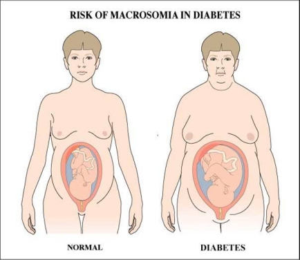
2. Diabetes
Next to macrosomia, the factor most closely associated with shoulder dystocia is maternal diabetes in pregnancy. The prevalence of diabetes in pregnant women is increasing due to an older pregnant population, a higher rate of obesity, and more thorough antenatal detection (Young 2013).
One of the first clear-cut demonstrations of this was Acker's 1985 paper showing the following:
|
Estimated fetal wt. |
Nondiabetic mothers
% shoulder dystocia |
Diabetic mothers
% shoulder dystocia |
| < 4000 g
|
1.1% |
3.7% |
| 4000-4499 g |
10.0% |
30.6% |
| > 4500 g |
22.6% |
50% |
As can be seen, babies of diabetic mothers had a three to fourfold increase in the risk of shoulder dystocia compared to babies of nondiabetic mothers in each weight category.
Although diabetic mothers accounted for only 1.4% of the birth population in this study, they accounted for 4.9% of shoulder dystocias. Acker also showed that although the general rate of Erb palsy following shoulder dystocia is roughly 10%, 17% of babies born to diabetic mothers developed Erb palsy.
Other investigators have shown similar or larger correlations between diabetes and shoulder dystocia:
Sandmire (1988) found a relative risk for shoulder dystocia in the babies of diabetic mothers of 6.5 compared to nondiabetic mothers.
In Al-Najashi's 1989 study, the rate of shoulder dystocia in babies weighing over 4000gm born of diabetic mothers was 15.7%. Babies born to nondiabetic mothers had a shoulder dystocia rate of 1.6%.
Casey (1997), in a study of over 62,000 patients, found the shoulder dystocia rate in his general obstetrical population to be 0.9% while in his patients with gestational diabetes it was 3%.
Tsur (2011), in a study from Israel, showed that the odds ratio for shoulder dystocia with diabetes was 1.8 compared to nondiabetic mothers.
Overland (2013), in a population of just under 2 million deliveries in Norway, reported that shoulder dystocia occurred in 0.73% percent of all deliveries but 3.95% of deliveries in which the mother had diabetes.
Mehta (2014): Diabetes increases the overall risk of shoulder dystocia by more than 70%. In his study, the incidence of macrosomia was 21% among diabetic mothers versus 7.6% among those who were nondiabetic. Gram for gram, the incidence of shoulder dystocia and injury is higher in diabetic mothers.
Hansen (2014) reported that in his patient population the ratio of shoulder dystocia of nondiabetic to diabetic mothers was 0.6%:1.9%, a 201% increase.
There are two main reasons for this correlation between diabetes and shoulder dystocia. In the first place, diabetes in pregnancy is strongly linked to macrosomia. The growth of babies of diabetic mothers represents not only their genetic potential for growth but also reflects the conversion to fat of the excess glucose substrates present in both mother and baby. Secondly, as previously mentioned, growth is not as evenly distributed between the head and trunk in the babies of diabetic mothers as it is in those of nondiabetic mothers. Rather, babies of diabetic mothers show a pattern of greater shoulder, chest, and abdominal growth. As Ellis summarized in 1982:
The infant of a diabetic mother has a different body configuration than the infant of a nondiabetic mother. Increased deposition of fat in various organs may be due to increased insulin secretion in response to hyperglycemia.
Can shoulder dystocia be predicted in babies of diabetic mothers?
In the 1980s several authors published studies purporting to show that they could predict which babies of diabetic mothers would be at high risk for shoulder dystocia.
Elliott (1982) claimed that by evaluating the chest and biparietal diameters in infants of diabetic mothers weighing more than 4000 g, he could reduce the incidence of traumatic morbidity at delivery from 27% to 9%.
Tamura (1986) found that in diabetic women fetal abdominal circumference values greater than the 90th percentile correctly predicted macrosomia in 78% of cases. In his study, when both the abdominal circumference and the estimated fetal weight exceeded the 90th percentile in pregnant women with diabetes, macrosomia was correctly diagnosed 88.8% of the time.
Mintz, in a promising study from 1989, published data showing that in his hands a combination of fetal abdominal circumference greater than the 90th percentile for gestational age and shoulder soft tissue width greater than 12 mm was the best predictor of macrosomia. His data reported a sensitivity of 96%, specificity of 89%, and "accuracy" — positive predictive value — of 93%. He also found a significant correlation between shoulder width and a high HgA1C (a blood test that measures blood sugar control over the preceding three months).
Unfortunately, these results have not been supported or replicated by other investigators. Multiple experts in the field of shoulder dystocia have published data from very large series that contradict the conclusions listed above. In addition, the results of the above studies are not as powerful as might first be assumed.
In Elliott's study, for instance, although he was able to show that a large number of babies meeting certain chest-biparietal diameter criteria were macrosomic, 39% of babies with these same parameters — chest/biparietal diameter ratio of > 1.4 — were not larger than 4000 g. In Tamura's study, although he was able to predict macrosomia in babies meeting certain abdominal circumference criteria, he still was unable to identify the vast bulk of macrosomic fetuses. As for Mintz's study, no one has yet been able to duplicate his results.
In fact, most studies have found that neither macrosomia nor shoulder dystocia can be reliably predicted in the babies of diabetic mothers.
Acker (1985) showed that by using the criteria of large baby and diabetic mother he could predict 54.7% of shoulder dystocias — but would miss 45.3% of them (false negatives).
Delpapa (1991) stated that the predictive value of estimated fetal weight in babies of diabetic mothers for predicting shoulder dystocia was not sufficiently accurate to reliably identify them.
Moreover, most diabetic mothers do not have macrosomic babies and the overwhelming majority of macrosomic infants are not babies of diabetic mothers.
There are two other studies of interest relating to this question.
Coen (1980) showed that although HgbA1C is a good marker for long-term monitoring of blood sugars in diabetic patients, it is not a good predictor of large-for-gestational age infants. The average HgA1C in mothers of large-for-gestational age infants in his study was 6.7; for mothers delivering normal sized babies the average HgA1C was 6.5 — too close to be clinically useful.
Casey (1997) reported that although the rate of shoulder dystocia was in fact increased in mothers with gestational diabetes, this was not manifest in an increase in the rate of Erb palsy.
The bottom line is that macrosomia is as difficult to predict in diabetic mothers as it is in the nondiabetic population.
� 3. Maternal weight gain
The data linking maternal weight gain and fetal birth weight are controversial.
Abrams (1995) and Langhoff-Roos (1987) both showed that total maternal weight gain was significantly correlated with infant birth weight.
Dawes (1991), however, was not able to confirm this:
There was no apparent correlation between maternal weight gain and birth weight between women giving birth to average for gestation or large for gestational age infants
Several other investigators have reported conflicting information as to the effect of patterns of maternal weight gain on ultimate fetal weight. Some studies have found second trimester weight gain to be the major determinate whereas others have found that the weight gain in the last trimester was the most important factor. Given the contradictory and confusing data on this subject, Dawes' closing statement is probably the most apt:
The variations in total (maternal) weight gain and incremental weight gain are so wide that these measurements are unlikely to be clinically useful.
4. Fetal sex
There is little data correlating fetal sex with macrosomia and shoulder dystocia. Although on average male babies do weigh slightly more than females, there is no data showing a significantly higher number of macrosomic male infants than female infants.
Resnick in his classic 1980 paper mentions fetal sex as a potential factor but does not supply data to substantiate his claim.
El Madany (1990) showed that 59.2% of babies experiencing shoulder dystocia in his study were male — statistically significant but not of much value as a clinical predictor.
� 5. Post-dates
Even though fetal growth slows in the last several weeks of pregnancy, there is still some growth as long as pregnancy continues. Thus the longer the baby remains in utero, the larger the baby will be — and the greater the risk of shoulder dystocia. Acker (1985) was one of the first to demonstrate this association. Chervenak confirmed this in 1989 when he reported that 25.5% of babies delivering at 41 weeks gestation were macrosomic while only 6% prior to 41 weeks were (risk ratio 4.2) in a group delivering between 38 and 40 weeks gestation. Hernandez (1990), too, found a direct correlation between post-date babies and an increased risk of shoulder dystocia. He attributed this entirely to the increased tendency of post-date babies to be macrosomic.
Overton in 2013 looked at this question in greater detail. He found that without correcting for weight, the rate of shoulder dystocia at 36 weeks is 27% of that at 40 weeks. However, after correcting for birth weight, the relative risk of shoulder dystocia at 36 weeks — compared to 40 weeks — was 1.68. Thus after adjustment for birth weight his results showed that there was a consistent reduction in the risk of shoulder dystocia from 36 weeks onward. This finding was particularly pronounced in pregnancies complicated by maternal diabetes.
Summary of antepartum risk factors
- Macrosomia and maternal diabetes are the main risk factors for shoulder dystocia
- Predicting fetal weight is extremely unreliable
- Other factors — maternal weight gain, fetal sex, and post dates — are secondary risk factors. They are correlated with an increased risk for shoulder dystocia but are only relevant to the degree that they increase the risk of fetal macrosomia
- Since multiparity increases the number of precipitous labors it may be a slight primary risk factor for shoulder dystocia
� C. Intrapartum risk factors
Various characteristics of labor and delivery have been claimed to be useful in predicting whether or not a given mother-baby pair will end up with a shoulder dystocia and possible brachial plexus injury.
1. Instrumental delivery
Several studies have clearly shown that labors that end in instrumental vaginal deliveries — vacuum or forceps — show a higher rate of shoulder dystocia in each fetal weight group.
Nesbitit (1998), for example, reported the following data:
|
Weight (g) |
SD % in unassisted births |
SD % in instrumental
deliveries |
| 4000-4250 |
8.4% |
12.2% |
| 4250-4500 |
12.3% |
16.7% |
| 4500-4750 |
19.9% |
27.3% |
| >4750 |
23.5% |
34.8% |
Baskett (1995) similarly showed a tenfold increase of shoulder dystocia and a fivefold increase in brachial plexus injury (BPI) with mid-forceps deliveries
| |
SD |
BPI |
|
SVD |
0.3% |
0.04% |
|
Low forceps deliveries |
0.9% |
0.06% |
|
Midforceps delivery |
2.8% |
0.5% |
Benedetti (1978) reported that in deliveries with the combination of a prolonged second stage of labor and a mid-pelvic delivery there was a 4.6% rate of shoulder dystocia -- compared to 0.4% when there was neither a prolonged second stage nor a mid pelvic delivery.
McFarland (1986) showed that the relative risk of brachial plexus injury was 18.3 for midforceps deliveries and 17.2 for vacuum deliveries when compared to unassisted vaginal deliveries.
Hansen in 2014 reviewed the literature on shoulder dystocia with assisted vaginal deliveries. He found a shoulder dystocia rate of 0.6% with spontaneous deliveries but a rate of 2.0% with operative vaginally deliveries, a relative difference of 254%.
Parantainen (2014), in a Finnish study of 42,964 deliveries with 152 shoulder dystocias, found a relative risk of 3.98 between spontaneous deliveries and vacuum assisted deliveries.
Mehta (2014) found that shoulder dystocias increased by 35 to 45% in vacuum and forceps-assisted deliveries. For nondiabetic mothers with assisted deliveries this translated to shoulder dystocia rates of 8.6% for infants weighing 4000 to 4250 g, 12.9% for infants weighing 4250 to 4500 g, 23% 4500 to 4750 g, and 29% for infants 4750 to 5000 g. The total adjusted odds ratio for shoulder dystocia with instrumentally assisted deliveries was 1.9.
Zuarez-Easton (2015) reached a similar conclusion; he found an OR of 3.6 between spontaneous and vacuum assisted deliveries in which there was a brachial plexus injury.
Is there a difference between the use of forceps or vacuum when it comes to increasing the risk of shoulder dystocia?
Bofill (1997) found that there was a non-significantly higher incidence of shoulder dystocia with vacuum assist versus forceps: 4.6% versus 1.9%.
Dall’Asta (2016), on the other hand, showed no difference in the rate of shoulder dystocia between the use of vacuum and forceps. He postulated that the use of the vacuum or forceps to expedite fetal head delivery may interfere with the spontaneous mechanism of rotation of the trunk and ultimately with the descent of the shoulders in the birth canal. The lack of difference in his study between forceps and vacuum, as compared to Bofill’s study, may perhaps be attributed to the implementation of safer vacuum equipment since 1997.
Thus it is clear that deliveries requiring instrumental assistance have a higher risk of shoulder dystocia and brachial plexus injury than do spontaneous vaginal deliveries. It is not clear, however, that it is the instrumental deliveries themselves that are to blame for these shoulder dystocias. It may well be that the mother's inability to push the baby out without assistance is due to fetal macrosomia, an altered distribution of fat between the fetal head, chest, shoulders, and abdomen, or descent of the shoulders in the A-P as opposed to an oblique orientation-- themselves major risk factors for shoulder dystocia.
2. Experience of the deliverer
Since the safe resolution of a shoulder dystocia involves specific obstetrical maneuvers and since shoulder dystocias occur relatively infrequently, it would seem that more experienced practitioners would have better outcomes in these situations merely by virtue of having seen more of them. Such an opinion would surely be voiced by most obstetricians and experienced labor and delivery nurses. However the data does not support this belief.
Acker in 1988 looked at the experience of the deliverer in relation to neonatal injuries following shoulder dystocia deliveries. He found that the number of Erb palsies following shoulder dystocias did not vary with either the number of years a physician had been in practice or the number of deliveries that physician performed. As Acker stated,
Most clinicians hardly gain expertise and confidence in the difficult manipulations required to resolve shoulder dystocia due to the rarity of the condition.
3. Labor abnormalities
Several studies have shown a higher incidence of shoulder dystocia in labors in which the second stage of labor is prolonged. Nevertheless -- and paradoxically -- shoulder dystocias are not infrequently seen during labors with very rapid second stages.
Hopewood (1982) found that there was a deceleration phase of active labor between eight to 10 cm in 58% of shoulder dystocia deliveries.
Acker (1985) showed that arrest disorders significantly increase the chance of shoulder dystocia with larger babies.
Gross (1987) showed that a prolonged deceleration phase and long second stage contributed to brachial plexus injury risk but that these were only weak predictors.
Al-Natasha (1989) found that both a delay in the second stage of labor and slowed descent of the fetal head in obese multiparous women greatly increased the possibility that a shoulder dystocia would occur.
Weizsaecker (2007) found that brachial plexus injury is often but not always preceded by dysfunctional labor. In his study, active phase abnormalities predominated among the mothers who experienced a shoulder dystocia. The most important risk factor for shoulder dystocia was a long deceleration phase. This increased the adjusted odds of brachial plexus injury almost 6 fold.
Tsur (2011) showed that the odds ratio for shoulder dystocia with slow rate of descent during the second stage of labor was 2.4.
But the literature has sometimes contradicted itself on this issue.
Acker, in that same 1985 article referenced above, states:
No particular labor abnormality was predictive of an increased incidence of shoulder dystocia relative to that encountered with a normal labor pattern, a spontaneous delivery, or both.
McFarland (1975) likewise reported the same rate of labor abnormalities of the active phase of labor and of the second stage of labor in both shoulder dystocia and control groups. He concluded that labor abnormalities could not serve as clinical predictors for the subsequent development of shoulder dystocia.
Hernandez (1990) reported that although there is a relationship between the length of various stages of labor and shoulder dystocia, 70% of patients who experienced shoulder dystocia had normal labor patterns.
Lurie (1995) found no correlation between the length of the stages of labor and shoulder dystocia. He showed that there was no difference in (1) the mean rate of dilatation, (2) the percentage of protracted labors, or (3) the mean duration of the second stage of labor in a group of mothers who experienced shoulder dystocia deliveries versus a group that delivered without complication. His conclusion was that protracted labor did not seem to be a risk factor for shoulder dystocia. As he says in his paper,
One could not identify shoulder dystocia in advance while analyzing the rate of cervical dilation or duration of the second stage of labor.
Even if disorders of labor were found to be correlated with shoulder dystocia, it is not clear whether this would represent an independent risk factor. It might merely confirm that labor disorders are more common with macrosomic babies and that macrosomic babies more commonly experience shoulder dystocia. To date there has been no major study evaluating the length of various stages of labor broken down by neonatal weight categories in relationship to shoulder dystocia deliveries.
To further complicate the issue, it is well known—as discussed above--that shoulder dystocias and brachial plexus injuries are often seen with short second stages of labor:
Acker (1988) found that 31.8% of all babies with Erb palsy were born after precipitate second stages of labor. As he explains,
The rapidity of descent may prohibit the fetal shoulders from entering the inlet in an oblique diameter, preclude adequate preparation for delivery, and add to nerve root trauma.
Gonen (2000) reported that 7 of 17 patients (41%) with brachial plexus injury had second stages of labor shorter than 10 minutes
� 4. Oxytocin and anesthesia
There does not appear to be any independent correlation between the use of either oxytocin or anesthesia and shoulder dystocia deliveries.
Oxytocin is generally used to increase the strength of uterine contractions. To the extent that oxytocin has to be used more frequently with macrosomic infants, it might have a secondary correlation with shoulder dystocia deliveries. But there is no published data linking oxytocin use with the incidence of shoulder dystocia independent of fetal weight.
Likewise with anesthesia; there is no reported increase in shoulder dystocia deliveries in labors in which conduction anesthesia is employed.
5. Episiotomy
There is no statistically significant relationship between the absence of episiotomy, the frequency of shoulder dystocia, and any subsequent neonatal injury. That this is the case is perplexing given that almost all protocols for the resolution of shoulder dystocia advocate making a "generous episiotomy". This recommendation appears to be without support in the obstetrical literature.
Gurewitsch (2004) demonstrated that management of severe shoulder dystocia with an episiotomy versus fetal manipulation alone or both does not influence neonatal depression rates.
Paris (2011) reviewed a total of 94,842 births in which there were 953 shoulder dystocias and 102 brachial plexus injuries. The rate of episiotomy with shoulder dystocia dropped from 40% in 1999 to 4% in 2009 with no change in the rate of brachial plexus injury per 1000 vaginally births.
Sagi-Dain in a 2015 review of 14 articles on the subject found no evidence supporting the use of episiotomy in the prevention and management of shoulder dystocia.
There are two possible reasons one might make an episiotomy in the case of a shoulder dystocia.
The first would be to make more room for the baby to emerge. In this situation the indications for making an episiotomy would be the same as in any delivery: alleviating soft tissue dystocia of the perineum. If the perineal tissue were tight, then an episiotomy might be helpful in delivering the baby. However, if the soft tissue of the vagina and vulva is pliable and stretches easily, as in most multiparous women, then an episiotomy will not make it any easier to free the anterior shoulder from behind the pubic bone.
The second possible indication for an episiotomy during a shoulder dystocia would be to allow more room for the obstetrician's hand to reach inside the vagina in order to perform rotational maneuvers or to attempt to deliver the baby's posterior arm. An episiotomy might be helpful in accomplishing these maneuvers in a woman whose perineal tissues impede access to the fetal shoulders. However, in a woman in whom the perineal tissues are lax enough to allow these maneuvers to be performed, the automatic making of an episiotomy will not facilitate delivery and would be unnecessary.
Thus the almost universal recommendation that an episiotomy be made during all shoulder dystocia deliveries is without literature or data support.
Combination of risk factors
As would be expected, several studies have shown that a combination of risk factors significantly increases the risk of shoulder dystocia.
Benedetti in 1978 published an article noting that the combination of macrosomia greater than 4000 g, prolonged second stage of labor, and mid pelvic operative vaginally delivery led to a 21% incidence of shoulder dystocia and a high rate of neonatal injury.
Mehta (2014) noted that in the setting of fetal macrosomia and a second stage of labor greater than two hours, performance of assisted vaginally delivery led to an increase rate of shoulder dystocia.
Busoni found that the combination of birth weight greater than 4000 g and vacuum delivery led to an odds ratio of 13.7 for shoulder dystocia; for birth weight over 4500 g with use of the vacuum the odds ratio was 21.5.
The greatest risk for shoulder dystocia occurs in those groups of women who have multiple risk factors. An obese woman with a large pregnancy weight gain and gestational diabetes will have a significantly greater likelihood of having a macrosomic baby and shoulder dystocia than will a woman who has just one of these risk factors. The worst possible combination of risk factors would be an obese mother with diabetes, an estimated large-for-gestational-age fetus, a prolonged second stage of labor, and a forceps delivery. The rate of shoulder dystocia in such a situation would approach 40%. So, Can shoulder dystocia and brachial plexus injury be predicted?
There are some authors who have always felt that shoulder dystocia can be prevented. O'Leary, in his book on shoulder dystocia, states:
A well-prepared obstetrician or midwife can anticipate this problem [shoulder dystocia] as a result of routinely identifying those risk factors that predispose to shoulder dystocia. Thus prevention requires identification of risk factors, which leads to anticipation of the problem . . . Identification of critical risk factors will lead to anticipation, which in turn will lead to prevention.
O'Leary then boldly goes on to say:
The presence of macrosomia of 4500 g alone is justification for cesarean section in nonobese women. The presence of macrosomia of 4000-4500 g may in itself be sufficient to warrant abdominal delivery when other risk factors, especially a platypoid (flat) pelvis, diabetes and/or obesity, are present.
But despite the certitude of his statements, O'Leary presents no data to support his recommendations.
Other authors have also tried to articulate guidelines for avoiding shoulder dystocia. Anchor (1988) has said:
We advocate the abdominal mode delivery for infants of diabetic gravidas whose best estimated fetal weight exceeds 4000 g.
Langer (1991) stated that if all infants of diabetic mothers who weighed 4250 g or more were delivered by cesarean section, the overall cesarean section rate would increase by only 0.26% while shoulder dystocia would be reduced by 76%. He goes on to acknowledge, however, that in the nondiabetic group there is no weight that provides an optimal threshold for cesarean section to avoid shoulder dystocia.
But statements such as these have represented the fringe of obstetrical opinion. It has been the consensus of the vast majority of obstetricians who have studied the subject that there is no real way to figure out which babies are likely enough to have shoulder dystocia to warrant changes in the management of their labors.
The basic issue is this: One can suspect shoulder dystocia all one wants. But is there some combination of factors that predicts shoulder dystocia with an accuracy great enough to make doing cesarean sections, performing early inductions, or making other changes in management a reasonable course of action? The answer by most experts in the field of shoulder dystocia has been "No." Certainly there are risk factors which do increase the odds of shoulder dystocia and brachial plexus injury occurring. But so many babies with each of these risk factors do not encounter shoulder dystocia and brachial plexus injury that it is difficult to justify changes in management of all labors on the basis of these suspicions.
The majority of studies in the obstetrical literature have not been able to show that the sensitivity or positive predictive value of various methods for predicting shoulder dystocia is high enough to justify interventions--which usually means cesarean section. While macrosomia, diabetes, prolonged second stage of labor, instrumental delivery, and other factors do indicate a statistically increased risk of having a shoulder dystocia, their low positive predictive value and high false positive rate make them clinically useless as tools for predicting -- and hence trying to prevent -- shoulder dystocia.
The entire issue is best summed up in Practice Bulletin #40 "Shoulder Dystocia" (2002, reaffirmed 2015) by the American College of Obstetricians and Gynecologists. They find the preponderance of current evidence consistent with the following positions:
Most cases of shoulder dystocia cannot be predicted or prevented because there are no accurate methods to identify which fetuses will develop this complication.
Ultrasonic measurement to estimate macrosomia has limited accuracy
Planned cesarean section based on suspected macrosomia is not a reasonable strategy
Planned cesarean section may be reasonable for the nondiabetic with an estimated fetal weight exceeding 5000 g or the diabetic whose fetus is estimated over 4500 g
Supporting the position of the American College of Obstetricians and Gynecologists on the lack predictability of shoulder dystocia are the thoughts of various shoulder dystocia investigators
Resnick (1980): Most babies with shoulder dystocia do not have risk factors. "The diagnosis will often be made only after delivery of the fetal head."
Acker (1986): Almost half (47.6%) of all shoulder dysoticas occurred in babies weighing less than 4000 g.
Al-Najashi (1989) 41% of shoulder dystocia deliveries in his series occurred in infants of average birth weight, that is 2500 to 3999 g.
Basket (1995): The profile of risk for shoulder dystocia -- prolonged pregnancy, prolonged second stage of labor, macrosomia, and assisted mid-pelvic delivery -- was not clinically useful because "the large majority of cases of shoulder dystocia occur in patients without these risk factors"
Rouse and Owen (1996) used a theoretical model involving performing cesarean section in women with suspected macrosomic fetuses in order to prevent permanent brachial plexus injury. They defined macrosomia as 4500 g. Their model predicted that 3695 cesarean sections would be needed to prevent one case of permanent injury.
Eckert (1997): The greatest number of injuries occurred in nondiabetic patients with birth weights of less than 4000 g.
Lewis (1998): Only 25% of shoulder dystocia cases had at least 1 significant risk factor . . . the positive predictive value of pre-partum risk factors for shoulder dystocia is less than 2% individually, 3% when combined.
Irion, in a 1998 Cochrane Systematic Review, indicated that there was no benefit in terms of improved maternal or infant outcomes associated with the induction of labor for suspected fetal macrosomia. He also noted that cesarean sections are not without risk. He calculated that if in a given country an additional 10,000 cesarean sections were performed in an attempt to prevent shoulder dystocia, there would likely be 900 severe postpartum hemorrhages, the need for 100 blood transfusions, and 600 each of wound infections, endometritis, and urinary tract infections. There also would likely be an additional 30 cesarean hysterectomies 35 women with venous thromboembolism, 30 women with severe morbidity requiring admission to intensive care, one hundred women with uterine rupture in a subsequent pregnancy were vagina birth to be attempted, and at least one additional maternal death.
Gonen (2000) showed that 740 cesarean deliveries were needed to prevent a single case of permanent neurologic damage if all mothers suspected to have fetuses weighing greater than 4500 g underwent elective cesarean section.
Gherman (2002): "Most of these preconception and prenatal risk factors have extremely poor positive predictive values and therefore do not allow the obstetrician to accurately and reliably predict the occurrence of shoulder dystocia."
Chauhan (2004): Due to inaccuracies in predicting fetal weights, among uncomplicated pregnancies suspicion of macrosomia is not an indication for induction or for primary cesarean section.
Cunningham, author of Williams Obstetrics (22nd edition, 2005) reports that 99.5% of babies weighing 4000-4500 gms had a safe vaginal delivery without shoulder dystocia.
Backe (2008) evaluated 30,574 births, 91 of which were diagnosed with brachial plexus injury, 15 of which were permanent. Although he identified various risk factors--shoulder dystocia, macrosomia, diabetes, vacuum extraction, and forceps delivery—their predictive power was poor. He concludes that “plexus injury is not well predicted by known risk factors”.
Nath (2012): 233 out of 241 patients treated at Texas nerve and paralysis Institute for brachial plexus palsy had shoulder dystocia at delivery. 80% of the patients in the study were not macrosomic. Instrumental use was 41%. Higher birth weight does not affect the prognosis of brachial plexus injury.
Dodd in 2012 showed that while there are a number of factors associated with an increased risk of shoulder dystocia, none are of sufficient sensitivity or positive predictive value to allow their use clinically to reliably and accurately identify the occurrence of shoulder dystocia. While they did find that maternal diabetes, induction of labor, and infant birthweight greater than 4000 g was associated with an increased risk of shoulder dystocia, they are poorly predictive of shoulder dystocia at a population level. The findings of the study reinforce the occurrence of shoulder dystocia as in “unpredictable and unpreventable obstetric emergency”. He notes that the Royal College of Obstetricians and Gynecologists does not recommend cesarean delivery for the prevention of shoulder dystocia.
Parantainen (2014) estimated that at least 30 unnecessary cesarean sections would be required to prevent one shoulder dystocia when using an optimal cut off for the most accurate ultrasound parameters for estimating fetal weight: abdominal diameter minus biparietal diameter of greater than 25 mm. Moreover, in his series the birth weight for shoulder dystocia babies was less than 4000 g in 35% of cases.
Peleg’s (2015) institution had a policy of counseling women about risks when there was a sonographically estimated fetal weight of greater than 4000 g. Their study was unable to show that a policy of elective cesarean for macrosomia significantly reduced the incidence of either shoulder dystocia or brachial plexus injury.
Palatnik (2016) attempted to predict the occurrence of shoulder dystocia prior to assisted vaginal deliveries by identifying significant risk factors and combining them into a prediction model. These factors included multiparity, maternal diabetes, chorioamnionitis, arrest of labor or maternal exhaustion, use of vacuum, and an estimated fetal weight of greater than 4000 g. Unfortunately, this model did not allow the accurate prediction of shoulder dystocia. The area under the receiver operating characteristic curve was 0.73, demonstrating only a modest ability to predict shoulder dystocia before performing an operative vaginally delivery.
Sentilhes (2016) discussed the guidelines for clinical practice of the French College of Gynecologist and Obstetricians. These guidelines state that according to the literature, only two characteristics are independent risk factors for shoulder dystocia: a history of a previous shoulder dystocia (which multiplies the risk by 10 – 20) and fetal macrosomia (risk multiplied by 6 – 20). Diabetes and maternal obesity are also consistently associated in the literature with an increased risk of shoulder dystocia (on the order of 2-4 times higher). But these associations are explained, at least in part, by the macrosomia they induce. The existence of a direct effect of maternal diabetes or obesity on this risk, independently of fetal weight, remains to be demonstrated. Nonetheless, even the factors associated continually and independently with shoulder dystocia do not enable its reliable prediction because they are not sufficiently discriminant. From 50 to 75% of all cases of shoulder dystocia occur in their absence, and the vast majority of deliveries in which they are present not involve it. Shoulder dystocia therefore remains an unpredictable obstetrical emergency.
As mentioned above, however, these conclusions may soon have to be changed. As the studies by the Hamilton group have shown, evaluation of multiple factors related to shoulder dystocia by means of a carefully researched algorithm seem to be able to predict with an impressive degree of certainty those fetuses at risk for shoulder dystocia and brachial plexus injury at the cost of no increase in the cesarean section rate.
next >>
Copyright © 2017 Henry Lerner
|